
QVI Case of the Month! For past cases,
click here
Patient is an interesting 84 y/o female who presents with a one month history of bilateral lower extremity swelling, varicose veins and tenderness. According to the patient, the swelling is marginally gravitational with the right leg worse than the left. She denies any previous history of DVT, SVT or malignancy. She also denies history of cardiac or arterial disease. She does have a history of hypertension and angina.
The right and left leg pulses are easily palpable at the femoral and popliteal; however ankle pulses are diminished bilaterally. No hair is noted on the toes and great toe capillary refill time appears slightly delayed bilaterally. Significant swelling is noted below the level of the knee bilaterally.
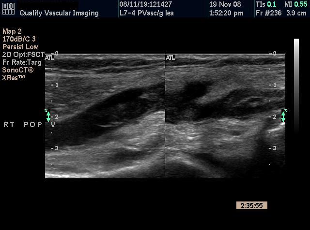
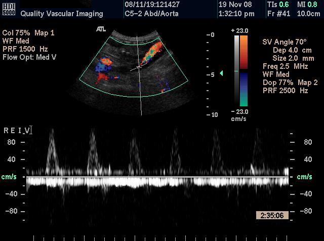
Ultrasound Findings : There is clear evidence of venous thrombosis in the right popliteal vein as well as the posterior tibial and peroneal veins. There was also thrombus noted in the small saphenous vein bilaterally. However, there was not sufficient evidence of infrainguinal venous obstruction that would explain the patients bilateral symptoms. Therefore, the examination was extended into the abdomen to evaluate the inferior vena cava (IVC) and iliac veins.
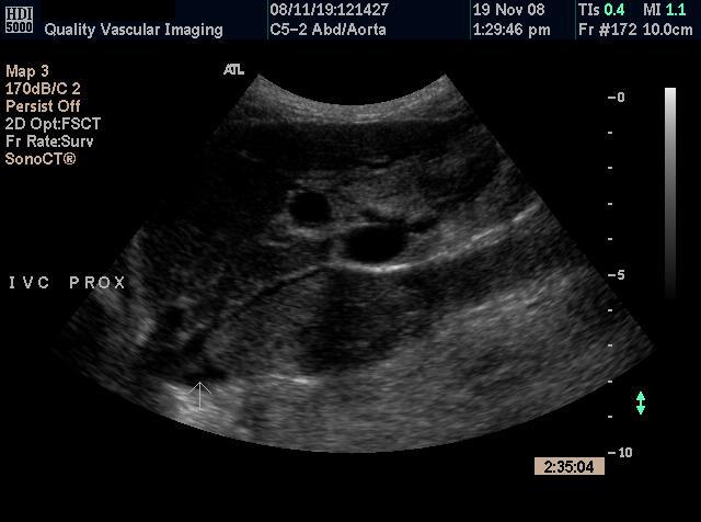
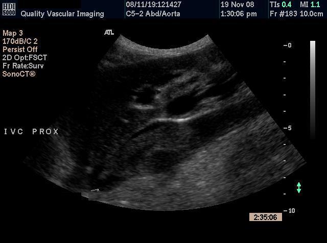
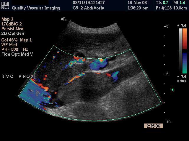
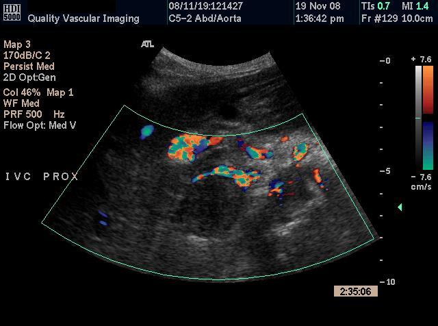
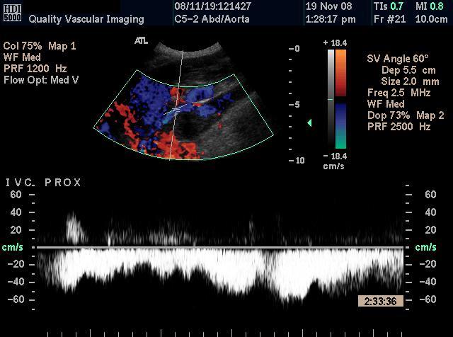
Proximal IVC Proximal IVC
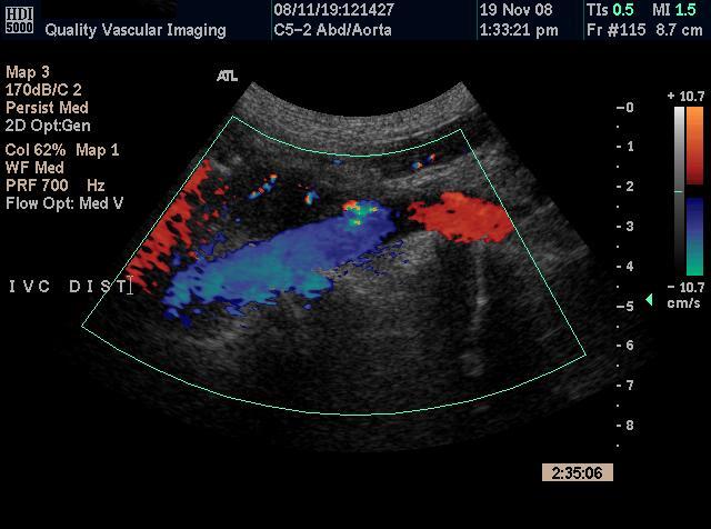
Proximal IVC showing flow around obstruction long (left) and cross section (right)
Spectral analysis of the flow in the proximal IVC (left) and the iliac vein (right) These very high quality images demonstrate near occlusion of the inferior vena cava. Given the infrainguinal thrombus, IVC thrombosis could be a distinct possibility. This however is not the case and the differentiation is made largely by the presentation, ie: no iliac involvement. Tumor should always be considered in isolated IVC obstruction.
Subsequent Findings: The patient was admitted to the emergency department and anticoagulants were started; however there was no placement of a Greenfield filter due to the fact that the inferior vena cava diameter exceeds the 2.8 centimeter limitation.
Computed Axial Tomography (CAT) exams were order of the chest, abdomen and pelvis with results as dictated by the interpreting radiologist follows:
Discussion: In general the incidence of thombosis of the inferior vena cava (IVC) is relatively low compared to the incidence of deep vein thombosis (DVT). Exact incidence of IVC thombosis (IVCT) is unknown due to the variability in the clinical presentations. According to WebMD:
Tumors Numerous malignancies have been associated with IVCT. Perhaps the most common is renal cell carcinoma. The intravascular tumor extends from the renal vein and can propagate as far as the heart. The tumor can partially or completely occlude the IVC. Not all intravascular irregularities of the kidney represent tumor either. One case has been reported of a patient who underwent radical nephrectomy for presumed renal cell carcinoma and was subsequently found to have only renal vein thrombosis. Other genitourinary tumors that reportedly cause IVCT include seminomas and teratomas. Numerous other less common tumors reportedly involve the IVC. Intuitively, any structure that is anatomically related to the IVC can generate either direct compression or vascular invasion. Retroperitoneal leiomyosarcoma, adrenal cortical carcinoma, and renal angiomyolipoma have all been reported as presenting in association with IVCT. Even hepatic hemangioma has caused IVCT from extrinsic compression. Additionally, malignancy itself is a risk factor for DVT and thus represents a risk factor for the extension of DVT into the IVC.
For past cases,
click here |
QVI Home Virtual Vein Center Why QVI really is the Best in Vascular Ultrasound! Case of the Month Patients Referring Physicians Health Professionals Site Map |

Current Happenings

Introducing our new educational website.
![]()
Virtual Vein Center is a new concept in educational delivery. Get the education you need and want, when you need it. If you need CME, you can get them here as well.
To read more about it, click here for a complete page. Feel free to go to the site and browse around.

Several QVI staff took time to attend the 2014 American College of Phlebology Annual Congress in Phoenix Arizona in November to deliver numerous workshops and lectures. It was a high quality meeting as usual. The complete program is available for download here.
The 2014 SVU Annual Conference was held in Orlando and several QVI attended and presented numerous presentations. Jeannie was also honored as a Fellow of the SVU.
To read more
Jeannie recently attended the 25th Society of Vascular Medicine 2014 Annual Conference as an invited speaker in La Jolla, Ca. Her numerous lectures were very well received.
The International Union of Phlebology, in conjunction with the American College of Phlebology held its World Meeting in Boston in September 2013. Held only every 4 years, this was the first time ever in the US. Several QVI staff were invited speakers presenting some original scientific research.
Sydney, Australia
Bill was the International Keynote Speaker at the Australian Sonographer Association Annual National Conference in Sydney.
What a great experience!
To read more about this and our other international teaching
QVI was once again awarded the D.E. Strandness Award for Scientific Excellence at the 2013 SVU Annual Conference.
To read more -
Medical Compression socks continue to be on the forefront of venous treatment. Recently, they have entered the realm of the athlete. To learn more about what compression socks can do you you, please visit compressionsocks.pro
QVI wins the D.E Strandness Award at the 2012 SVU Annual Conference!
Read more about it!
To go to the
CASE OF THE MONTH!
Click the QVI logo
Compression Socks?
We carry the BEST!
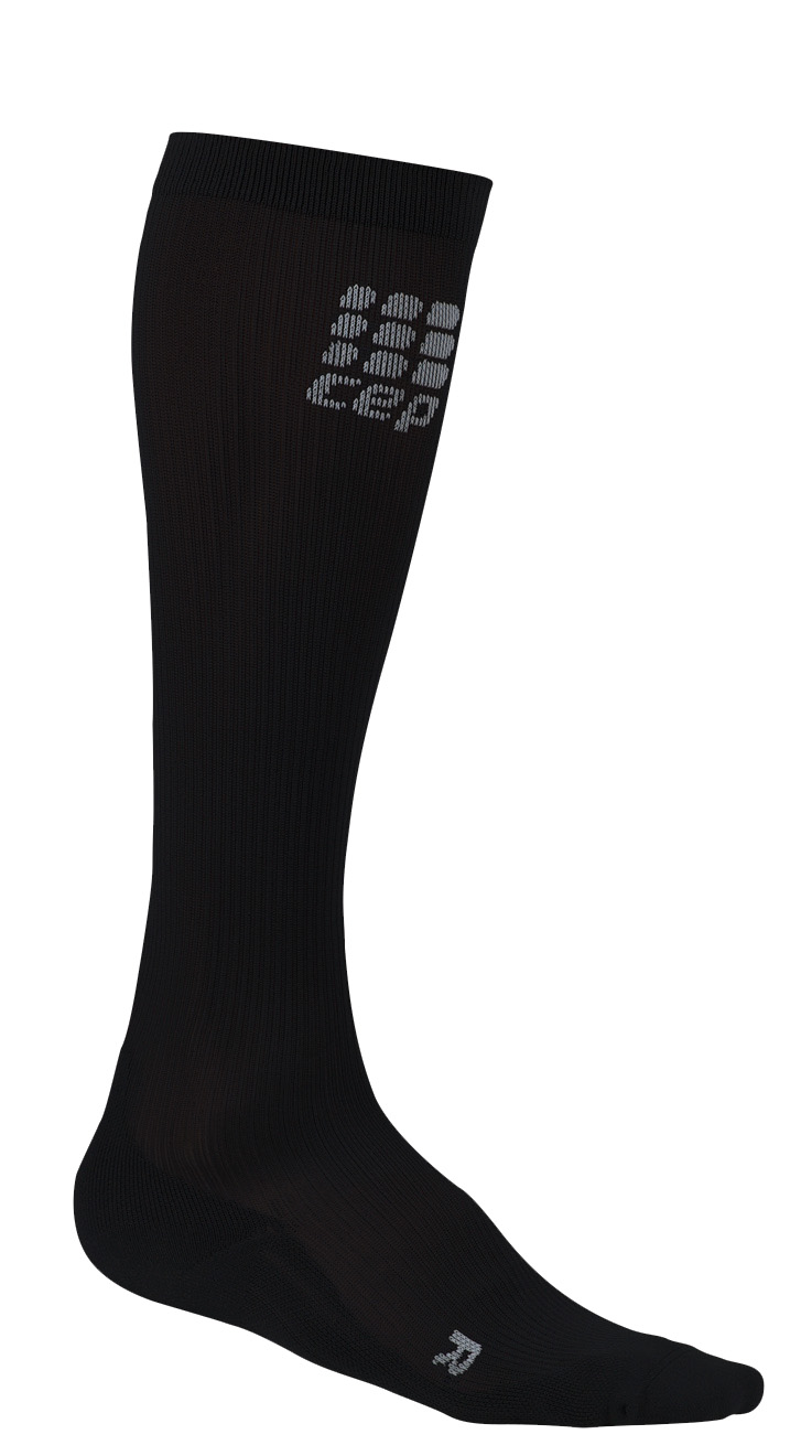
Compression socks are not only a main stay in the treatment of venous and lymphatic disease but are now used by elite athletes everywhere. Please visit our webstore for more information about compression therapy by clicking the image above! .